Immunoglobulin A
Immunoglobulin A (Ig A, also referred to as sIgA in its secretory form) is an antibody that plays a role in the immune function of mucous membranes.The oligomeric forms of IgA in the external (mucosal) secretions also contain a polypeptide of a much larger molecular mass (70 kDa) called the secretory component that is produced by epithelial cells.This molecule originates from the poly-Ig receptor (130 kDa) that is responsible for the uptake and transcellular transport of J chain-containing polymeric (but not monomeric, which is devoid of J chain) IgA across the epithelial cells and into secretions such as tears, saliva, sweat and gut fluid.[citation needed] In the blood, IgA interacts with an Fc receptor called FcαRI (or CD89), which is expressed on immune effector cells, to initiate inflammatory reactions.[15] Ligation of FcαRI by IgA containing immune complexes causes antibody-dependent cell-mediated cytotoxicity (ADCC), degranulation of eosinophils and basophils, phagocytosis by monocytes, macrophages, and neutrophils, and triggering of respiratory burst activity by polymorphonuclear leukocytes.[15] Polymeric IgA (mainly the secretory dimer) is produced by plasma cells in the lamina propria adjacent to mucosal surfaces.[19] Immune exclusion is a process of agglutinating polyvalent antigens or pathogens by crosslinking them with antibody, trapping them in the mucus layer, and/or clearing them peristaltically.[19] Since sIgA is a poor opsonin and activator of complement, simply binding a pathogen isn't necessarily enough to contain it—specific epitopes may have to be bound to sterically hinder access to the epithelium.Additionally, Blastocystis species have been shown to have several subtypes that generate cysteine and aspartic protease enzymes which degrade human IgA.

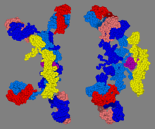
H-chainL-chainJ-chainsecretory componentantibodyimmune functionmucous membranesimmunoglobulinmucous secretionssalivacolostrumgenitourinary tractgastrointestinal tractprostaterespiratory epitheliummicrobescomplement systemopsonizesisotypesglycosylateddisulfidenon-covalentgut-associated lymphoid tissueB-cell receptorPolymersmonomersJ chainpolypeptidecysteinemolecular weightepithelial cellsFc receptorantibody-dependent cell-mediated cytotoxicityeosinophilsbasophilsphagocytosismonocytesmacrophagesneutrophilspolymorphonuclear leukocytesalternativelectin pathwaysplasma cellspolymeric immunoglobulin receptorlamina propriabasolateralendocytosisluminal surfaceProteolysisM cellsdendritic cellsclass switchingmesenteric lymph nodesepithelialpolyvalentperistalticallysterically hinderasialoglycoprotein receptorsgalactoseglycansselective IgA deficiencyimmunodeficiencyNeisseriaNeisseria gonorrhoeaegonorrheaStreptococcus pneumoniaeHaemophilus influenzaeprotease that destroys IgABlastocystisaspartic proteaseIgA nephropathyCeliac diseaseHenoch–Schönlein purpuracomplement component 3VancomycinList of target antigens in pemphigusTGF betaFăgărășan S.BibcodeMedical Subject HeadingsLymphocyticadaptive immune systemcomplementAntigenSuperantigenAllergenAntigenic variationHaptenEpitopeLinearConformationalMimotopeAntigen presentationprofessional APCsDendritic cellMacrophageB cellImmunogenMonoclonal antibodiesPolyclonal antibodiesAutoantibodyMicroantibodyPolyclonal B cell responseAllotypeIsotypeIdiotypeImmune complexParatopeImmunityAutoimmunityAlloimmunityAllergyHypersensitivityInflammationCross-reactivityCo-stimulationToleranceCentralPeripheralClonal anergyClonal deletionTolerance in pregnancyImmune privilegeImmunogeneticsAffinity maturationSomatic hypermutation