Endocytosis
[3] More recent experiments have suggested that these morphological descriptions of endocytic events may be inadequate, and a more appropriate method of classification may be based upon whether particular pathways are dependent on clathrin and dynamin.The principal components of the endocytic pathway are:[3] It was recently found that an eisosome serves as a portal of endocytosis in yeast.In endocytosis, the clathrin coat is assembled on the cytoplasmic face of the plasma membrane, forming pits that invaginate to pinch off (scission) and become free CCVs.The actual budding-in process, whereby a pit is converted to a vesicle, is carried out by clathrin; Assisted by a set of cytoplasmic proteins, which includes dynamin and adaptors such as adaptin.Coated pits and vesicles were first seen in thin sections of tissue in the electron microscope by Thomas F Roth and Keith R.[27] The importance of them for the clearance of LDL from blood was discovered by Richard G. Anderson, Michael S. Brown and Joseph L. Goldstein in 1977.Recent studies have also discovered that polymerase I, transcript release factor, and serum deprivation protein response also play a role in the assembly of caveolae.[31] The process of cell uptake depends on the tilt and chirality of constituent molecules to induce membrane budding.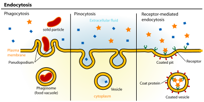

cellular processsubstancescell membranevesiclepinocytosisphagocytosisDe DuveÉlie Metchnikoffsynaptic vesiclemembranesreceptor-mediated endocytosiscaveolaeClathrin-mediated endocytosiscytosolicclathrinClathrin-coated vesiclesreceptorslow density lipoproteintransferringrowth factorsantibodiescaveolincholesterolglycolipidssmooth musclepneumocytesfibroblastsadipocytesendothelial cellsPotocytosiscytosolendosomeslysosomesmicroorganismsapoptoticdynaminmacropinocytosisphagosomesmannose-6-phosphateGolgi apparatuselectron microscopyvacuoleseisosomeplasma membraneAP2 adaptorsLDL receptoradaptinKeith R. PorterMichael S. BrownJoseph L. GoldsteinBarbara PearseoligomersGolgi complexblood-brain barrierα-helicesSARS-CoV-2epithelial cellActive transportEmperipolesisExocytosisTrans-endocytosisBibcodeMembrane transportbiological membranesPassive transportSimple diffusionmediated transportFacilitated diffusionOsmosisChannelsCarriersUniporterSymporterAntiporterPrimary active transportSecondary active transportCytosisEfferocytosisNon-specific, adsorptive pinocytosisTranscytosisDegranulation