Cytotoxic T cell
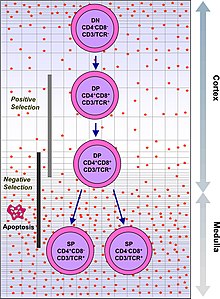
An antigen is a molecule capable of stimulating an immune response and is often produced by cancer cells, viruses, bacteria or intracellular signals.However, CD8+ T cells also have the ability to make some cytokines, such as TNF-α and IFN-γ, with antitumour and antimicrobial effects.In the first place, naïve (virgin) T-lymphocytes are an initial stage of T cell residing in the thymus which have not yet encountered an antigen with affinity for its TCR.In the thymus-independent pathway, because the APC is infected, it is highly activated and expresses a large number of co-receptors for coactivation.If activation occurs, the lymphocyte polarizes its granules towards the site of the synapse and releases them, producing a "lethal hit".Through the action of perforin, granzymes enter the cytoplasm of the target cell and their serine protease function triggers the caspase cascade, which is a series of cysteine proteases that eventually lead to apoptosis (programmed cell death).[13] Due to high lipid order and negatively charged phosphatidylserine present in their plasma membrane, TC cells are resistant to the effects of their perforin and granzyme cytotoxins.[19] Platelets have been shown to facilitate the accumulation of virus-specific cytotoxic T cells into the infected liver.In different studies, rheumatoid arthritis is strongly linked to major histocompatibility complex (MHC) class II antigens.Activated CD4+ T lymphocytes stimulate monocytes, macrophages and synovial fibroblasts to elaborate the cytokines interleukin-1, interleukin-6 and tumour necrosis factor alpha (TNFa), and to secrete metalloproteinases.Moreover, several animal studies suggest that cytotoxic T cells may have a predominantly proinflammatory effect in the disease.For example, HIV has adopted very high mutation rates to allow them to escape recognition by CD8+ T cells.[26] Studies in a diabetic mouse model showed that CD4+ cells are responsible for the massive infiltration of mononuclear leukocytes into pancreatic islets.However, in studies with NOD mice carrying a null mutation at the beta-2 microglobulin (B2M) locus and thus lacking major histocompatibility complex class I molecules and CD8+ T cells, it was found that they did not develop diabetes.Cytotoxic T-lymphocytes have been implicated in the development of various diseases and disorders, for example in transplant rejection (cytotoxic T-lymphocytes attack the new organ after detecting it as foreign, due to HLA variation between donor and recipient);[30] in excessive cytokine production in severe SARS-CoV-2 infection (due to an exaggerated lymphocyte response, a large amount of pro-inflammatory cytokines are generated, damaging the subject);[31][32] inflammatory and degenerative diseases of the central nervous system, such as multiple sclerosis (T cells become sensitised to certain proteins, such as myelin, attacking healthy cells and recruiting more immune cells, aggravating the disease).
Natural killer T cell"helper" CD4+ cellsT lymphocytewhite blood cellcancerpathogensvirusesbacteriaT-cell receptorsantigenimmune responsecancer cellsclass I MHCglycoproteinaffinitycytokinesTNF-αIFN-γthymusiodinestressHematopoietic stem cellsbone marrowV(D)J recombinationproteinT cellsgamma delta T cellschemokineshuman cytomegalovirusco-receptorsantigensapoptosisautoimmunityCD4+ T cellsnucleatederythrocytesintracellularpathogenantigen processingT cell antigen receptorantigen-presenting cellMHC class IcostimulatorsT helper cellsdendritic cellsmemory T cellshelper T cellsinterleukin 2differentiationsomatic cellsperforingranzymesgranulysinserine proteasecaspaseFAS ligandT lymphocyteslamin Apoly ADP ribose polymeraseDNA-PKcsEomesoderminantibodiesbacterialhepatitis B virusPlateletsCXCR5+CXCL13arthritissynovial membranehyperplasiaantigen-presenting cellsinterleukin-1interleukin-6tumour necrosis factor alphacomplementType 1 diabetespancreatic isletsbeta-2 microglobulinchemotherapy-induced peripheral neuropathytransplant rejectionSARS-CoV-2pro-inflammatory cytokinesmultiple sclerosismyelinCTL-mediated cytotoxicityList of distinct cell types in the adult human bodyBibcodeLymphocytesB cellsB1 cellPlasmablastPlasmaMemoryFollicularMarginal zoneNaïveBreg cellB10 cellTransitional B cellThymocyteHelper CD4+RegulatoryMemory T cellInnate-like T cellsMucosal associated invariant T cellInnate lymphoid cellsNK cellsCytokine-induced killer cellLymphokine-activated killer cellNull cellAdaptive NK cellUterine natural killer cellsType 1 innate lymphoid cellsType 2 innate lymphoid cellsNuocytesType 3 innate lymphoid cellsLTi cellsLymphopoiesisHematopoietic stem cellLymphoblastProlymphocyteLymphocyticadaptive immune systemSuperantigenAllergenAntigenic variation