Dorsal column–medial lemniscus pathway
At the level of the medulla oblongata, the fibers of the tracts decussate and are continued in the medial lemniscus, on to the thalamus and relayed from there through the internal capsule and transmitted to the somatosensory cortex.[2] The DCML pathway is made up of the axons of first, second, and third-order sensory neurons, beginning in the dorsal root ganglia.The gracile fasciculus carries sensory information from the lower half of the body entering the spinal cord at the lumbar level.The column reaches the junction between the spinal cord and the medulla oblongata, where lower body axons in the gracile fasciculus connect (synapse) with neurons in the gracile nucleus, and upper body axons in the cuneate fasciculus synapse with neurons in the cuneate nucleus.[6] First-order neurons secrete substance P in the dorsal horn as a chemical mediator of pain signaling.It also allows for the ability known as haptic perception (stereognosis), to determine what an unknown object is, using the hands without visual or audio input.Fine touch is detected by cutaneous receptors called tactile corpuscles that lie in the dermis of the skin close to the epidermis.Damage to the dorsal column-medial lemniscus pathway below the crossing point of its fibers results in loss of vibration and joint sense (proprioception) on the same side of the body as the lesion.Damage above the crossing point result a loss of vibration and joint sense on the opposite side of the body to the lesion.


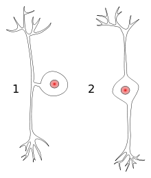
sensory receptorsproprioceptivePrecursorNeural tubeSystemSomatosensory systemDecussationMedial lemniscusSensorimotor cortexAnatomical terms of neuroanatomysensory pathwaycentral nervous systemsensationsfine touchvibrationtwo-point discriminationproprioceptionsomatosensory cortexpostcentral gyrusparietal lobegracile fasciculuscuneate fasciculusdorsal columnsfuniculimedulla oblongatadecussatethalamusinternal capsulespinal cordbrainstemfirst-order neuronssecond-order neuronsthird-order neuronssensory neuronsdorsal root gangliaafferent fibersmake contact withdorsal column nucleigracile nucleuscuneate nucleusmedullaventral posterolateral nucleuscervicallumbarinternal arcuate fiberssensory decussationmyelinatedventral nuclear groupsixth thoracic vertebrafirst-ordersecond-orderpseudounipolar neuroncell bodybipolar neuronaction potentialmechanoreceptortissuepseudounipolardorsal root ganglionposterior rootposterior hornposterior columnfuniculusposterolateralposterior median sulcusglial cellssynapsesubstance Plower limbupper limbtrigeminal nerveventral posteromedial nucleusventral posterior nucleusprimary somatosensory cortexhumanshaptic perceptioncutaneous receptorstactile corpusclesdermisepidermismuscle spindlesmechanoreceptorsMerkel cellsbulbous corpuscleslamellar corpusclesdendritesame sideopposite sideRomberg's testBrown-Séquard syndromeKarl Friedrich BurdachFriedrich GollSpinothalamic tractU AlbertaCervical enlargementLumbar enlargementConus medullarisFilum terminaleCauda equinaMeningesCentral canalTerminal ventricleGrey columnsPosterior grey columnMarginal nucleusSubstantia gelatinosa of RolandoNucleus propriusRexed lamina VRexed lamina VILateral grey columnIntermediolateral nucleusPosterior thoracic nucleusAnterior grey columnInterneuronAlpha motor neuronOnuf's nucleusGamma motor neuronRexed laminaeCentral gelatinous substanceGray commissureWhite matterPosteriorPosterior column-medial lemniscus pathwayGracileCuneateLateral