Electron microscope
[2] One significant step was the work of Hertz in 1883[3] who made a cathode-ray tube with electrostatic and magnetic deflection, demonstrating manipulation of the direction of an electron beam.In 1931, Max Knoll and Ernst Ruska[12][13] successfully generated magnified images of mesh grids placed over an anode aperture.He stated in a very brief article in 1932[16] that Siemens had been working on this for some years before the patents were filed in 1932, claiming that his effort was parallel to the university development.[23] Although current transmission electron microscopes are capable of two million times magnification, as scientific instruments they remain similar but with improved optics.[26] By the early 1980s improvements in mechanical stability as well as the use of higher accelerating voltages enabled imaging of materials at the atomic scale.[27][28] In the 1980s, the field emission gun became common for electron microscopes, improving the image quality due to the additional coherence and lower chromatic aberrations.The choice of workflow will be highly dependent on the application and the requirements of the corresponding scientific questions, such as resolution, volume, nature of the target molecule, etc.Another example is high resolution mass spectrometry (ion microscopy), which has been used to provide correlative information about subcellular antibiotic localisation,[59] data that would be difficult to obtain by other means.[60] An early example of these ‘volume EM’ workflows was simply to stack TEM images of serial sections cut through a sample.The next development was virtual reconstruction of a thick section (200-500 nm) volume by backprojection of a set of images taken at different tilt angles - TEM tomography.More recently, back scattered electron (BSE) images can be acquired of a larger series of sections collected on silicon wafers, known as SEM array tomography.The increased volume available in these methods has expanded the capability of electron microscopy to address new questions,[60] such as mapping neural connectivity in the brain,[65] and membrane contact sites between organelles.Microscopes designed to achieve high resolutions must be housed in stable buildings (sometimes underground) with special services such as magnetic field canceling systems.Samples of hydrated materials, including almost all biological specimens, have to be prepared in various ways to stabilize them, reduce their thickness (ultrathin sectioning) and increase their electron optical contrast (staining).


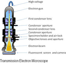


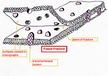
Scanning tunneling microscopemicroscopeelectronselectron opticselectron diffractionresolutionlight microscopesTransmission electron microscopyScanning transmission electron microscopyScanning electron microscopeElectron microprobeLow-energy electron microscopyPhotoemission electron microscopyErnst RuskaEmil WiechertArthur WehneltHans BuschDennis GaborLeó SzilárdTechnische HochschuleTechnische Universität BerlinMax KnollNobel prizeSiemens-SchuckertReinhold RüdenbergBodo von BorriesHelmut RuskaManfred von ArdenneWashington State UniversityUniversity of TorontoEli Franklin BurtonJames HillierAlbert CreweUniversity of Chicagofield emission sourcefield emission gunTransmission electron microscopehigh voltageelectron beamelectron gunelectromagneticdetectorphosphorscintillatorzinc sulfidefibre opticdigital cameraspherical aberrationhigh-resolution transmission electron microscopyangstrompicometresannular dark-field imagingBacillus subtilisraster scanninglow-energy secondary electronscathodoluminescenceMicroanalysisX-ray crystallographycoated in goldscanning electron microscope (SEM)sputter-coatedUltramicrotomyStainingCryofixationChemical millingSputteringfixationproteinsaldehydesformaldehydeglutaraldehydelipidsosmium tetroxidevitreous (non-crystalline) iceethanecryo-electron microscopyfocused ion beamethanolacetonecritical point dryingresinsfreeze dryingpropylene oxideAralditeDurcupanacrylic resinstainedhypochloritegalliumNegative stainnanoparticlesuranyl acetatephosphotungstic acidultramicrotomediamondglass knivesuraniumtungstengreyscalevacuumliquid-phase electron microscopyenvironmental scanning electron microscopein situ electron microscopycarbon nanotubesdiatomcryofixedList of materials analysis methodsElectron energy loss spectroscopyEnergy filtered transmission electron microscopyImmune electron microscopyMicroscope image processingMicroscopyNanotechnologyScanning confocal electron microscopyThin sectionTransmission Electron Aberration-Corrected MicroscopeBibcodeCowley JMWayback MachineElectron microscopyHistoryMicrographTimeline of microscope technologyElectronAuger effectBremsstrahlung