Endosome
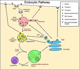
Endosomes can be classified as early, sorting, or late depending on their stage post internalization.LDL dissociates because of the slightly acidified environment of the early endosome, generated by a vacuolar membrane proton pump V-ATPase.Phosphatidyl inositol phosphates (PIPs), one of the most important lipid signaling molecules, is found to differ as the endosomes mature from early to late.The inter-conversion of these lipids is a result of the concerted action of phosphoinositide kinases and phosphatases that are strategically localized[17] There are three main compartments that have pathways that connect with endosomes.Also, in some circumstances, late endosomes/MVBs fuse with the plasma membrane instead of with lysosomes, releasing the lumenal vesicles, now called exosomes, into the extracellular medium.There is no consensus as to the exact nature of these pathways, and the sequential route taken by any given cargo in any given situation will tend to be a matter of debate.[18] In the opposite direction, retromer generates vesicles at early endosomes that carry molecules back to the Golgi.Molecules that follow these pathways include the receptors for LDL, epidermal growth factor (EGF), and the iron transport protein transferrin.The activated EGFRs stimulate their own ubiquitination, and this directs them to lumenal vesicles (see below) and so they are not recycled to the plasma membrane.[26] Molecules that follow these pathways include LDL and the lysosomal hydrolases delivered by mannose-6-phosphate receptors.Also, the transmembrane EGFRs, bound to EGF, are tagged with ubiquitin and are therefore sorted into lumenal vesicles by the ESCRTs.
VorarephiliaHeLa cellsorganelleseukaryoticendocytictrans Golgi networkplasma membranelysosomescell membraneendocytic cycleGolgi apparatusendomembrane systemlow-density lipoproteinLDL receptorV-ATPaseepidermal growth factormannose 6-phosphate receptorligandstransferrin receptorsTransferrinreceptormacrophagesPhagosomesmacropinosomesautophagosomesPI(4,5)P2plasma membranesPI(3)PPI(4)Pkinasesphosphatasesmelanocytesepithelialtranscytosisexosomesclathrin-coated vesicleretromerreceptor-mediated endocytosisclathrincaveolinreceptorsCholesterolEGF receptorTransmembrane proteinsubiquitinendosomal sorting complexes required for transportBack-FusionExosomeParamural bodyBibcodeNature Reviews Molecular Cell BiologyFrontiers in PharmacologyNucleusEndoplasmic reticulumParenthesomeAutophagosomeVesicleLysosomePhagosomeVacuoleAcrosomeCytoplasmic granuleMelanosomeMicrobodyGlyoxysomePeroxisomeWeibel–Palade bodyCytoskeletonMicrofilamentIntermediate filamentMicrotubuleProkaryotic cytoskeletonMicrotubule organizing centerCentrosomeCentrioleBasal bodySpindle pole bodyMyofibrilUndulipodiumCiliumAxonemeRadial spokePseudopodiumLamellipodiumFilopodiumEndosymbiontsMitochondrionPlastidChloroplastChromoplastGerontoplastLeucoplastAmyloplastElaioplastProteinoplastTannosomeApicoplastNitroplastNucleolusRibosomeSpliceosomeCytoplasmCytosolInclusionsProteasomeMagnetosomeCell wallExtracellular matrix