Temporal lobe epilepsy
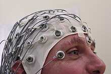
[10] These types of TLE are very rare due to the genetic cause or lesions such as tumor, birth defect, blood vessel abnormalities in the temporal lobe.[10][2] The common mesial temporal lobe seizure auras include a rising epigastric feeling, abdominal discomfort, taste (gustatory), smell (olfactory), tingling (somatosensory), fear, déjà vu, jamais vu, flushing, or rapid heart rate (tachycardia).[15][16][14]: 71 Mesial temporal lobe epilepsy arising from the language dominant hemisphere impairs verbal memory, and mesial temporal lobe epilepsy arising from the language non-dominant hemisphere impairs nonverbal memory.[19][20][21][22][23] Hippocampal sclerosis, brain tumor, traumatic brain injury, cerebral vascular malformation, neuronal migration disorders, infections such as encephalitis and meningitis, autoimmune disease (limbic encephalitis) and genetic disorders may cause temporal lobe epilepsy.[27] Experimental research has shown that N-methyl-d-aspartate (NMDA) receptor activation causes neuronal cell loss, and electrical stimulation-induced animal models of temporal lobe epilepsy duplicate the cell loss pattern of temporal lobe epilepsy in humans.[27] Repetitive seizures irreversibly damage interneurons leading to persistent loss of recurrent inhibition.[29]: 1318 In the normal brain, dentate granule cells block seizure spread from entorhinal cortex to the hippocampus.[3] Magnetoencephalography may diagnose temporal lobe epilepsy by recording epileptiform discharges or seizure patterns arising from the magnetic fields of neural electrical currents.[31] CT scan is advised in emergencies when the suspected cause of epilepsy may be intracerebral hemorrhage, brain abscess, large cerebral infarction or subdural empyema.CT scan may better demonstrate calcium containing brain abnormalities causing epilepsy such as in tuberous sclerosis and Sturge–Weber syndrome.[34]: 751 Studies show that language dominant anterior temporal lobectomy may lead to verbal memory decline.[16] However, study outcomes are more variable on language non-dominant anterior temporal lobectomy leading to nonverbal memory decline.[40] Remission was more likely among those without hippocampal sclerosis, brain tumor, or focal cortical dysplasia on MRI scan.


Lobes of the brainTemporal lobeSpecialtyNeurologyPsychiatrybrain disorderunprovoked seizuresepilepsymesial temporal lobelateral (neocortical) temporal lobeMemorycomorbiditieselectroencephalographicneuroimagingAnticonvulsant medicationsepilepsy surgerydietary treatmentsInternational League Against Epilepsyclassification of the epilepsiesbiological neural networkcerebral hemispheremedialhippocampusparahippocampal gyrusamygdalalateralepigastricdéjà vujamais vuflushingtachycardiaautomatismsopposite side of the braindysphasialanguage dominantvertigoparietal lobebilateral tonic-clonicdeclarative memoryepisodic memorysemantic memoryverbal memorynonverbal memorymajor depressive disorderpost-traumatic stress disordergeneral anxiety disorderpsychosisobsessive-compulsive disorderschizophreniabipolar disordersubstance use disordersuicideGeschwind syndromesexualityreligiosityHippocampal sclerosisbrain tumortraumatic brain injuryvascular malformationneuronal migration disordersencephalitismeningitisautoimmune diseaselimbic encephalitisgenetic disordersfebrile seizuresstatus epilepticusBrain positron emission tomographyN-methyl-d-aspartate (NMDA) receptoranimal models of temporal lobe epilepsyinterneuronsrecurrent inhibitionGABAergicneuronal firingglutamatergicprincipal neuronsexcitatorysubicularγ-Aminobutyric acidreceptorsneuronal depolarizationgranule cell dispersionentorhinal cortexmossy fiber pathwaypyramidal cellsFocal cortical dysplasiacortical layersdysmorphic neurons, balloon cellsglutamateVagus nerve stimulationResponsive neurostimulation devicedeep brain stimulationelectroencephalgramLong-term video-EEG monitoringMagnetoencephalographymagnetic resonance imagingcavernous hemangiomahypoxic-ischemic brain injuryglucosemetabolismSingle-photon emission computed tomographyComputed tomographyintracerebral hemorrhagebrain abscesscerebral infarctionsubdural empyemacardiac pacemakerdefibrillatorcochlear implanttuberous sclerosisSturge–Weber syndromeAnticonvulsantpharmacoresistantPenfieldanterior temporal lobectomyrandomized controlled trialstereotactic radiosurgerystereotactic radiofrequency ablationvagus nerve stimulatorvagus nerveketogenic dietmodified Atkins dietDiseasesDBMedlinePluseMedicinePatient UK