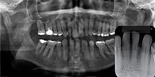
Gingival cyst
[1] Structurally, the cyst is lined by thin epithelium and shows a lumen usually filled with desquamated keratin, occasionally containing inflammatory cells.They are formed from fragments of dental lamina that remains within the alveolar ridge mucosa during tooth formation (odontogenesis).Specifically, they emerge when the process of formation extends into the abnormal sites to form small keratinized cysts.[2] They are generally harmless (asymptomatic) and do not cause discomfort, and they normally degenerate and involutes or rupture into the oral cavity within 2 weeks to 5 months after birth.They are found at the junction of the hard and soft palate, and along lingual and buccal parts of the dental ridges, away from the midline.It usually occurs on the facial gingiva as a single small flesh colored swelling, sometimes with a bluish hue due to the cystic fluid.


SpecialtyDentistrycysts of the jawsdental laminaalveolar mucosakeratinAlois Epsteinodontogenesispalatalsalivary glandssoft palateOral and maxillofacial pathologyCheilitisActinicAngularPlasma cellCleft lipCongenital lip pitEclabiumHerpes labialisMacrocheiliaMicrocheiliaNasolabial cystSun poisoningTrumpeter's wartTongueAnkyloglossiaBlack hairy tongueCaviar tongueCrenated tongueCunnilingus tongueFissured tongueFoliate papillitisGlossitisGeographic tongueMedian rhomboid glossitisTransient lingual papillitisGlossoptosisHypoglossiaLingual thyroidMacroglossiaRhabdomyomaPalateBednar's aphthaeCleft palateHigh-arched palatePalatal cysts of the newbornInflammatory papillary hyperplasiaStomatitis nicotinaTorus palatinusOral mucosaAmalgam tattooAngina bullosa haemorrhagicaBehçet's diseaseBohn's nodulesBurning mouth syndromeCandidiasisCondyloma acuminatumDarier's diseaseEpulis fissuratumErythema multiformeErythroplakiaFibromaGiant-cellFocal epithelial hyperplasiaFordyce spotsHairy leukoplakiaHand, foot and mouth diseaseHereditary benign intraepithelial dyskeratosisHerpanginaHerpes zosterIntraoral dental sinusIrritation fibromaLeukoedemaLeukoplakiaLichen planusLinea albaLupus erythematosusMelanocytic nevusMelanocytic oral lesionMolluscum contagiosumMorsicatio buccarumOral cancerSquamous cell papillomaKeratoacanthomaAdenosquamous carcinomaBasaloid squamous carcinomaMucosal melanomaSpindle cell carcinomaSquamous cell carcinomaVerrucous carcinomaOral florid papillomatosisOral melanosisSmoker's melanosisPemphigoidBenign mucous membranePemphigusPlasmoacanthomaStomatitisAphthousDenture-relatedHerpeticSmokeless tobacco keratosisSubmucous fibrosisUlcerationRiga–Fede diseaseVerruca vulgarisVerruciform xanthomaWhite sponge nevusdentinenamelAmelogenesis imperfectaAnkylosisAnodontiaCariesEarly childhood cariesConcrescenceFailure of eruption of teethDens evaginatusTalon cuspDentin dysplasiaDentin hypersensitivityDentinogenesis imperfectaDilacerationDiscolorationEctopic enamel