Cerebral organoid
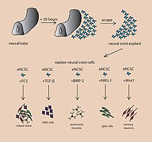
[1][2] The brain is an extremely complex system of heterogeneous tissues and consists of a diverse array of neurons and glial cells.This complexity has made studying the brain and understanding how it works a difficult task in neuroscience, especially when it comes to neurodevelopmental and neurodegenerative diseases.The varying physiology between human and other mammalian models limits the scope of animal studies in neurological disorders.Neural organoids contain several types of nerve cells and have anatomical features that recapitulate regions of the nervous system.Below is an image showing some of the chemical factors that can lead stem cells to differentiate into various neural tissues; a more in-depth table of generating specific organoid identity has been published.These organoids can be used in experiments that current in vitro methods are too simplistic for, while also being more applicable to humans than rodent or other mammalian models might be.Embryoid bodies are composed of three layers: endoderm, mesoderm and ectoderm, which has the potential to be differentiated into different types of tissue.Replication of specific brain regions in cerebral organoid counterparts is achieved by the addition of extracellular signals to the organoid environment during different stages of development; these signals were found to create change in cell differentiation patterns, thus leading to recapitulation of the desired brain region.[8] It has been shown that cerebral organoids grown using the spinning bioreactor 3D culture method differentiate into various neural tissue types, such as the optic cup, hippocampus, ventral parts of the teleencephelon and dorsal cortex.[1] Genetic markers for the hippocampus, ventral forebrain, and choroid plexus are also present in cerebral organoids, however, the overall structures of these regions have not yet been formed.[7] The cortical plate is usually generated inside-out such that later-born neurons migrate to the top superficial layers.[1] In DishBrain, grown human brain cells were integrated into digital systems to play a simulated Pong via electrophysiological stimulation and recording.[12][13][14] In the 2020s, significant changes in how these electrophysiological systems are made and interact with brain organoids could lead to better stimulation and recording data across the organoind in 3D.This suggests that local stimuli are released once one or more cells differentiate into a specific type as opposed to a random pathway throughout the tissue.[17] Cerebral organoids also provide a unique insight into the timing of development of neural tissues and can be used as a tool to study the differences across species.[25] Cerebral organoids can be used as simple models of complex brain tissues to study the effects of drugs and to screen them for initial safety and efficacy.[26][27][28] This can be used for the prevention and treatment of specific diseases[29] (see below) but also for other purposes such as insights into the genetic factors of recent brain evolution (or the origin of humans and evolved difference to other apes),[30][31][32] human enhancement and improving intelligence, identifying detrimental exposome impacts (and protection thereof), or improving brain health spans.Cerebral organoids have been used in studies in order to understand the process by which Zika virus affects the fetal brain and, in some cases, causes microcephaly.[20][21] Cerebral organoids infected with the Zika virus have been found to be smaller in size than their uninfected counterparts, which is reflective of fetal microcephaly.[20] In one case, a cerebral organoid grown from a patient with microcephaly demonstrated related symptoms and revealed that apparently, the cause is overly rapid development, followed by slower brain growth.The disease is difficult to reproduce in mouse models because mice lack the developmental stages for an enlarged cerebral cortex that humans have.[41] By cultivating cerebral organoids from ASD patients with macrocephaly, connections could be made between certain gene mutations and phenotypic expression.The significance of this use of brain organoids is that it has added great support for the excitatory/inhibitory imbalance hypothesis[43] which if proven true could help identify targets for drugs so that the condition could be treated.[44] Further research into the extent and accuracy by which cerebral organoids recapitulate epigenetic patterns found in primary samples is also needed.Cerebral organoid can be used to model prenatal pathophysiology and to compare the susceptibility of the different neural cell types to hypoxia during corticogenesis.Other limitations include: Until recently, the central part of organoids have been found to be necrotic due to oxygen as well as nutrients being unable to reach that innermost area.[19] The structure of cerebral organoids across different cultures has been found to be variable; a standardization procedure to ensure uniformity has yet to become common practice.[25] Ethical concerns have been raised with using cerebral organoids as a model for disease due to the potential of them experiencing sensations such as pain or having the ability to develop a consciousness.Steps are being taken towards resolving the grey area such as a 2018 symposium at Oxford University where experts in the field, philosophers and lawyers met to try to clear up the ethical concerns with the new technology.[51] Similarly, projects such as Brainstorm from Case Western University aim to observe the progress of the field by monitoring labs working with brain organoids to try to begin the ‘building of a philosophical framework’ that future guidelines and legislation could be built upon.


in vitroorganoidspluripotent stem cellsneuronsglial cellsin vivocortexretinaspinal cordthalamushippocampusStem cellsembryoidorganoidendodermmesodermectodermembryoid bodycell cultureneuroectodermmatrigelSMAD inhibitionbioreactorcell doubling timescerebral cortexoccipital lobeGenetic markersreelinCajal-Retzius cellsglutamatetetrodotoxininto digital systemsTranscriptome analysisTUNEL assaysCrispr/Cas 9Adeno-associated viruselectroporationAging brainNeurobiological effects of physical exerciseWetware computerMetabolomeNeurogenomicscell fate potentialcell replacement therapyTissue morphogenesisvertebratescell migrationNeural glial cellsfate mappingneurodegenerationvasculatureimmunologicallyhigh-throughput screeningevolvedhuman enhancementexposomeimproving brain health spansZika viruscytochrome P450CYP3A5microcephalymicrocephalinAlzheimer's diseaseplaquesamyloid beta proteinsneurofibrillary tanglesAutismGABAergicepigeneticsDNA methylationglioblastomastumorigenesisnecroticsynaptogenesisNeuroethicsconsciousnessHumanzeecontroversialNeural tissue engineeringBibcodeMuotri, Alysson R.