Visual memory
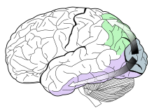
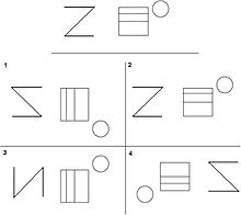
Visual memory describes the relationship between perceptual processing and the encoding, storage and retrieval of the resulting neural representations.A majority of experiments highlights a role of human posterior parietal cortex in visual working memory and attention.We therefore have to establish a clear separation of visual memory and attention from processes related to the planning of goal-directed motor behaviors.[4] Activity in the posterior parietal cortex is tightly correlated with the limited amount of scene information that can be stored in visual short-term memory.[4] These results suggest that the posterior parietal cortex is a key neural locus of our impoverished mental representation of the visual world.However, ample evidence indicates that object identity and location are preferentially processed in ventral (occipito-temporal) and dorsal (occipito-parietal) cortical visual streams, respectively.Although visual short term memory is essential for the execution of a wide array of perceptual and cognitive functions, and is supported by an extensive network of brain regions, its storage capacity is severely limited.[4] The retrieval of long term visual memories is associated with activation of both anterior and posterior temporal cortices.[9] The participants results from each task are then assessed and placed into six categories; omissions, distortions, preservations, rotations, misplacements, and sizing errors.With the use of brain imaging devices researchers able to further investigate memory performance above and beyond standard tests based on exact response times, and activation.[10] Subjects are blindfolded and instructed to lay motionless while simultaneously eliminating any visual imagery present in their mind's eye.[10] After the scan is complete a control has been formed which can be compared with activated regions of the brain while performing visual memory tasks.It is thought of as a three-dimensional cognitive map, which contains spatial features about where the person is and visual images of the area, or an object being concentrated on.A classic test of spatial memory is the Corsi block-tapping task, where an instructor taps a series of blocks in a random order and the participant attempts to imitate them.In a recent study where a visual search task was administered quiet rest or sleep is found to be necessary for increasing the amount of associations between configurations and target locations that can be learned within a day.[22] Studies have shown that with aging, in terms of short-term visual memory, viewing time and task complexity affect performance.In a recent study visual working memory and its neutral correlates was assessed in university students who partake in binge drinking, the intermittent consumption of large amounts of alcohol.This study looked at the neural correlated of the low level of response to alcohol using functional magnetic resonance imaging during a challenging visual memory task.The results were that young people who report having needed more alcohol to feel the effects showed higher levels of brain response during visual working memory, this suggests that the individual's capacity to adjust to cognitive processing decreases, they are less able to adjust cognitive processing to contextual demands."All of the hallucinatory palinopsia symptoms occur concomitantly in a patient with one lesion, which supports current evidence that objects, features, and scenes are all units of visual memory, perhaps at different levels of processing."[3] Studying the excitability alterations associated with palinopsia in migraineurs could provide insight on mechanisms of encoding visual memory.It has also been found in postmortem examinations of the brains of people with reading disabilities that they have fewer neurons and connections in the areas representing the transient visual systems.[29] However, there is debate over whether this is the only reason for reading disabilities, scotopic sensitivity syndrome, deficits in verbal memory and orthographic knowledge are other proposed factors.[30] These case studies show that these two types of visual memory are located in different parts of the brain and are somewhat unrelated in terms of functioning in daily life.


human eyeencodingstorageretrievalmind's eyepalinopsiastimulusvisual object recognitiontemporal cortexparietal cortexPosterior parietal cortexparietal lobeworking memoryposterior cortexconsolidationvisual cortexhemisphereOccipital lobelateral geniculate bodyDorsal streamVentral streamlimbic systemoccipital lobesVisual short-term memoryprefrontal cortexanterior cingulate cortexBenton Visual Retention Testvisual perceptionabilitiessensitivityreading disabilitiesnonverbal learning disabilitiestraumatic brain injuryattention-deficit disorderalzheimer'sdementiadistortionsrotationsscoresgendereducationinteractiongeometricalencoderecallneuroimagingneural networksmemoryBaddeley and Hitch's modelvisualencodedcognitive mapconcentratedmental imageimaginesretrievingEidetic memoryIconic memorymemory systemunconsciouslydecaysSpatial memoryhippocampusrandom orderimitateaveragevulnerableprocessingtextureorientationventral regionsinterferenceElizabeth Loftusmemory errorsacademicchalkboardChildrenBrain damageeffect of alcoholbinge drinkingfunctional magnetic resonance imagingHallucinatory palinopsiaseizureslettersvisual processingeye movementssynchronizationpostmortem examinationsbrainsneuronsscotopic sensitivity syndromediseasetrauma to the brainWayback MachineBibcodeMental imagescognitionSaksida, L.M.PsychologyHistoryPhilosophyPsychologistBasic psychologyAbnormalAffective neuroscienceAffective scienceBehavioral geneticsBehavioral neuroscienceBehaviorismCognitiveCognitivismCognitive neuroscienceSocialComparativeCross-culturalCulturalDevelopmentalDifferentialEcologicalEvolutionaryExperimentalGestaltIntelligenceMathematicalNeuropsychologyPerception