Unhappy triad
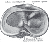
The strong valgus or rotary force to the knee tears the ACL, MCL, and medial meniscus all together.In about 10% of cases, the force is applied to the opposite side of the knee, and the lateral and posterolateral ligaments are torn.The medial meniscus is a C-wedge shaped piece of cartilage that acts as a"shock absorber" between the femur and the tibia.It is a fibrocartilage band on the lateral side of the knee joint and can easily be injured with torsional stress or direct force.Without them, the body weight is distributed unevenly on the femur and tibia, possibly leading to early arthritis in the knee joint.This poses a problem in a meniscus injury, as blood flow is diminished and the avascular areas tend to not heal.PT includes exercise ambulatory programs, mobilizations, and modalities to help ease symptoms and speed up the recovery process.The purpose of physical therapy is strengthening muscle and increasing the knee's range of motion without damaging the new grafts.Physical Therapist will provide immediate knee mobilization manually or with continuous passive motion (CPM) within the first week.59/100 patients were injured during contact sports, 30/100 in downhill skiing and 11/100 in other recreational activities, traffic accidents or at work.Non-weightbearing in the injury situation led to the same rate of MCL tears (18/28) as weightbearing (35/72) but significantly more intact menisci (19/28 vs 23/72).Thus, contact sports injuries were more often sustained during weightbearing, with a resultant joint compression of both femuro-tibial compartments as shown by the higher incidence of bicompartmental meniscal lesions.[13] In 1936, Cambell stated that an "impairment of the anterior crucial and medial ligaments is associated with injuries of the internal cartilage".A review of all arthroscopically confirmed acute injuries of second degree or worse to the ACL and MCL was performed.


SpecialtyEmergency medicineinjuryanterior cruciate ligamentmedial collateral ligamentmeniscuspatellaposterior cruciate ligamentlateral collateral ligamentvalgusmedial meniscuslateral meniscusanterior cruciate ligament injuryanterior superior iliac spineexternal semilunar fibrocartilagefibrocartilageavasculartear of meniscusautograftarthroscopicallyterrible triad of the elbowshoulderMacDonald triadAnterior cruciate ligament reconstructionMedlinePlusMedlinePlus EncyclopediaDislocationssubluxationssprainsstrainsJointsligamentsDislocation of jawWhiplashupper armDislocated shoulderSeparated shoulderALPSA lesionSLAP tearBankart lesionforearmPulled elbowGamekeeper's thumbHip dislocationPosterior cruciate ligament injuryPatellar dislocationKnee dislocationSprained ankleHigh ankle sprainTurf toeMusclestendonsRotator cuff tearPulled hamstringPatellar tendon ruptureAchilles tendon ruptureShin splints