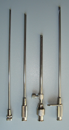
Thoracentesis
Thoracentesis /ˌθɔːrəsɪnˈtiːsɪs/, also known as thoracocentesis (from Greek θώραξ (thōrax, GEN thōrakos) 'chest, thorax' and κέντησις (kentēsis) 'pricking, puncture'), pleural tap, needle thoracostomy, or needle decompression (often used term), is an invasive medical procedure to remove fluid or air from the pleural space for diagnostic or therapeutic purposes.[9] Routine measurement of coagulation profiles is generally not indicated, however; when performed by an experienced operator "hemorrhagic complications are infrequent after ultrasound-guided thoracentesis, and attempting to correct an abnormal INR or platelet level before the procedure is unlikely to confer any benefit".[10] Relative contraindications include cases in which the site of insertion has known bullous emphysema, use of positive end-expiratory pressure (PEEP, see mechanical ventilation) and only one functioning lung (due to diminished reserve).Traditional expert opinion suggests that the aspiration should not exceed 1 L to avoid the possible development of pulmonary edema, but this recommendation is uncertain as the volume removed does not correlate well with this complication.[5] Major complications are pneumothorax (3–30%), hemopneumothorax, hemorrhage, hypotension (low blood pressure due to a vasovagal response) and reexpansion pulmonary edema.Exudate Transudate A high amylase level (twice the serum level or the absolute value is greater than 160 Somogy units) in the pleural fluid is indicative of either acute or chronic pancreatitis, pancreatic pseudocyst that has dissected or ruptured into the pleural space, cancer or esophageal rupture.
Chest X-raypleural effusionICD-9-CMMedlinePlusthoraxpleural spacecannulalocal anesthesiaMorrill WymanHenry Ingersoll Bowditchmidaxillary lineintercostal spacemedical emergencychest tubeeffusionscancercongestive heart failurepneumoniasurgerytuberculosispneumothoraxpleural fluidhemothoraxtube thoracostomycoagulationbullous emphysemapositive end-expiratory pressuremechanical ventilationhemopneumothoraxhemorrhagepulmonary edemahematomaseromadyspneaultrasoundetiologyLight's criteriatransudateexudateInfectionInflammationMalignancyIatrogenicConnective tissue diseaseEndocrine disorderspericarditisNephrotic syndromeHypoalbuminemiaCirrhosisAtelectasisdialysisSuperior vena cava obstructionpancreatitispseudocystGlucosedifferential diagnosisempyemaBoerhaave syndromeChylothoraxlymph vesselstriglyceridecholesterolchylous effusionserousthoracic ductlymphomawhite blood cellsred blood cellsmicrobiological cultureGram stainZiehl–Neelsen stainCytologylung cancermetastasispleural mesotheliomarespiratory systemUpper RTRhinoplastySeptoplastySomnoplastyAlarplastyRhinectomyRhinomanometryAcoustic rhinometrySinusotomylarynxLaryngoscopyLaryngectomyLaryngotomyThyrotomyLaryngotracheal reconstructionLower RTtracheaCricothyrotomyTracheoesophageal punctureTracheotomybronchusBronchoscopyPneumonectomyLobectomyWedge resectionTransplantationDecorticationHeart–lung transplantChest wallpleuramediastinumdiaphragmpleural cavityPleurodesisThoracoscopyThoracotomyMediastinoscopyNuss procedureMedical imagingBronchographyCT pulmonary angiogramHigh-resolution computed tomographySpiral CTVentilation/perfusion scanClinical prediction rulePneumonia severity indexCURB-65Lung function testBody plethysmographySpirometry