Glomerulus (kidney)
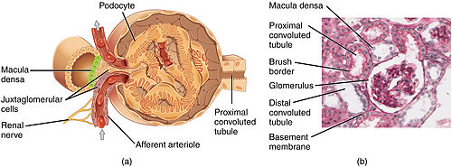
The walls have a unique structure: there are pores between the cells that allow water and soluble substances to exit and after passing through the glomerular basement membrane and between digitating podocyte foot processes, enter the capsule as ultrafiltrate.It consists mainly of laminins, type IV collagen, agrin, and nidogen, which are synthesized and secreted by both endothelial cells and podocytes.[4] The space between adjacent podocyte foot processes is spanned by slit diaphragms consisting of a mat of proteins, including podocin and nephrin.[3] This provides tighter control over the blood flow through the glomerulus, since arterioles dilate and constrict more readily than venules, owing to their thick circular smooth muscle layer (tunica media).These vasa recta run adjacent to the descending and ascending loop of Henle and participate in the maintenance of the medullary countercurrent exchange system.The main function of the glomerulus is to filter plasma to produce glomerular filtrate, which passes down the length of the nephron tubule to form urine.This arrangement of two arterioles in series determines the high hydrostatic pressure on glomerular capillaries, which is one of the forces that favor filtration to Bowman's capsule.The factors that influence permselectivity are the negative charge of the basement membrane and the podocytic epithelium, as well as the effective pore size of the glomerular wall (8 nm).[6] The rate of filtration from the glomerulus to Bowman's capsule is determined (as in systemic capillaries) by the Starling equation:[6] The walls of the afferent arteriole contain specialized smooth muscle cells that synthesize renin.About 175 years later, surgeon and anatomist William Bowman elucidated in detail the capillary architecture of the glomerulus and the continuity between its surrounding capsule and the proximal tubule.


B. Glomerular basement membrane: 1. lamina rara interna 2. lamina densa 3. lamina rara externa
C. Podocytes: 1. enzymatic and structural proteins 2. filtration slit 3. diaphragma

Bowman's capsuleproximal tubulePrecursorMetanephric blastemaNephronkidneyAnatomical terminologycapillariesintraglomerular mesangial cellsfiltrateafferent arterioleefferent arteriolesvenulesultrafiltrationrenal corpuscleglomerular filtration rateefferent arterioleendothelial cellsglomerular basement membranepodocyte foot processesfenestraeblood plasmared blood cellswhite blood cellsplateletspodocyteslamininscollagennidogenalbuminglobulinfoot processslit diaphragmspodocinnephringlycocalyxserum albuminpericytesbasement membraneefferentarteriolevenulesmooth muscletunica mediainterlobular veinrenal veinjuxtamedullary nephronsvasa rectarenal medullaloop of Henlecountercurrent exchangecollecting ductsrenal calyxplasmaarterioleshydrostatic pressureTable of permselectivity for different substancespermeabilitynegative chargesodiumpotassiumhemoglobinoncotic pressureStarling equationjuxtaglomerular cellsrenin–angiotensin systemblood volumepressureurinalysisdiabetic kidney diseaseglomerulonephritisglomerulosclerosisIgA nephropathynephrotoxicityMarcello MalpighiWilliam BowmanGlomerulusBlood–brain barrierScanning electron microscopeWayback Machineurinary systemKidneysFasciaCapsuleCortexcolumnMedullapyramidsmedullary interstitiumCortical lobuleMedullary rayCirculationRenal arterysegmentalinterlobararcuateinterlobularafferentPeritubular capillariesPodocyteFiltration slitsMesangiumIntraglomerular mesangial cellRenal tubuleProximal convoluted tubuleDescendingThin ascendingThick ascendingDistal convoluted tubuleCollecting duct systemConnecting tubulePapillary ductTubular fluidRenal papillaMinor calyxMajor calyxRenal pelvisJuxtaglomerular apparatusMacula densaExtraglomerular mesangial cellUretersUreteropelvic junctionBladderVesical arteriesVesical veinsVaginal artery